PhysiologyMaps Home > Cardiovascular main map

Cardiac Myocytes

The heart is comprised of cardiac myocytes, or muscle cells. These cells work together to achieve excitation-contraction coupling, so the heart can continuously pump blood out to the body (aka, maintain the cardiac cycle). However, some of these myocytes have specialized functions that we don't normally think of when we think of "muscle cells". There are three functionally distinct groups of cardiac myocytes:

  1. Nodal cells: Found at the sinoatrial (SA) node and atrioventricular (AV) node, and function to initiate and conduct electical signal through the heart tissue,
  2. Atrial and ventricular myocytes: Make up the main mass of the atria and ventricles, and function to initiate muscle contraction in response to electrical signal, and
  3. His-Purkinje system: Found in the interventricular septum and ventricular walls, and function to quickly spread electrical signal throughout the main mass of the ventricles.

PhysiologyMaps Home > Cardiovascular main > Cardiac myocytes

Electrical Excitation of the Heart

Electrical signal flows through the heart to trigger contraction of the heart muscle cells (cardiac myocytes). The flow of electrical signal is started at the top of the heart (by the nodal cells of the SA node, in the right atrium) and is passed along between neighboring cardiac myocytes going down to the bottom of the heart. It is often referred to as a “wave of depolarization”, because each heart cell has its own action potential and will depolarize when a neighboring cell depolarizes. As heart cells pass on electrical signal on to their neighboring cells, it causes this massive spread of depolarization down the heart (in a “wave”). The whole purpose of this wave of electrical excitation is to trigger muscle contraction by the cardiac myocytes, which causes the heart to beat and pump out blood.

General flow of electrical signal through the heart

It makes sense this electrical signal flows in the direction it does: We want the atria to contract first, in order to empty blood into the ventricles during diastole; then, after a pause at the AV node, we want the ventricles to contract, pushing blood out to the lungs and body during systole. Thus, the flow of electrical signal helps maintain the cardiac cycle, or the constant flow of floow through the heart and body.


PhysiologyMaps Home > Cardiovascular main > Cardiac myocytes

Nodal Cells

Nodal cells are specialized cardiac myocytes that initiate and conduct electrical signal in the heart at the beginning of each heartbeat. Nodal cells have the ability generate spontaneous action potentials (automaticity); therefore, nodal cells can initiate their own depolarizations and set the pace of the heart. Nodal cells do not contribute to generating force during heart muscle contraction, like atrial and ventricular myocytes do.

Nodal cells are found in two clusters in the heart:

  1. the sinoatrial (SA) node (main pacemaker of the heart) and
  2. the atrioventricular (AV) node (backup pacemaker of the heart).


PhysiologyMaps Home > Cardiovascular main > Cardiac myocytes

Sinoatrial (SA) Node

The sinoatrial (SA) node is a small cluster of conducting cells located in the top of the right atrium. The SA node's main function is to initiate the electrical signal for each heartbeat that travels through our atrial myocytes, to our AV node, and through our ventricular myocytes. The SA node is therefore considered the main "pacemaker" of the heart, because the SA node's firing rate sets our heart rate. The SA node can set the heart rate because of its inherent automaticity, or ability to generate its own action potentials. The SA node normally initiates every wave of depolarization through the heart, because of all the cardiac myocytes that can initiate their own depolarization, the SA node has the fastest firing rate (~60-90 times per minute, a normal resting heart rate).

SA Node Action Potentials

The SA node’s action potential is a result of the flow of electrical current--driven by the movements of various ions according to their equilibrium potentials--through SA nodal cells with each heartbeat. It resembles a neuronal action potential, but with a few key differences.

  • Upstroke (“phase 0”): L-type (and some T-type) Ca2+ channels open when enough Na+ has leaked into cell and membrane potential reaches firing threshold. Ca2+ flows into cell.
  • Repolarization (“phase 3”): K+ flows out of cell to bring membrane potential back its more polarized membrane potential (and closer to K+'s equilibrium potential) in response to rapid Ca2+ influx. K+ channels are basically always open, so this happens pretty easily.
  • Unstable resting membrane potential (spontaneous depolarization, “phase 4”): SA nodal cells do not have a stable “resting” membrane potential. This is because of Na+ channels on SA nodal cells called “funny channels”, which are constantly open and allowing a steady leak of Na+ into the cell. This allows the membrane potential of SA nodal cells to slowly approach their firing threshold, where Ca2+ channels open and cause the upstroke. (This is different from neurons or atrial and ventricular myocytes, which must wait for a neighboring cell to depolarize and spread its signal; neurons stay at a constant resting membrane potential until a nearby signal causes it to depolarize.) These funny channels (and this constant upward climb toward the threshold potential) are the key to SA nodal cells’ automaticity--they don’t have to wait for a nearby cell to trigger depolarization, they can just depolarize on their own.

Regulation of the SA Node

The SA node sets the heart rate, which is tightly regulated by the autonomic nervous system. The effects of the autonomic nervous system on SA node firing rate (and, therefore, heart rate) are called “chronotropic effects”.


PhysiologyMaps Home > Cardiovascular main map > Cardiac myocytes

Atrioventricular (AV) Node

The atrioventricular (AV) node is a small cluster of conducting cells located in the bottom of the right atrium near the interventricular septum. The AV node’s main function is to conduct electrical signal from the atria to the ventricles. After the atria depolarize, the electrical signal reaches the AV node, where there is a slight delay (due to the slow conduction velocity of the AV node). This delay allows time for the newly depolarized atria to contract and empty their blood into the ventricles (this is diastole, aka filling of the ventricles) before the AV node sends off the signal to depolarize the His-Purkinje and the ventricles.

AV Node Action Potentials

The AV node’s action potential very closely resembles the SA node’s action potential. The main differences are: 1.) the AV node has a slower pacemaker rate (less steep phase 4 depolarization) and 2.) the AV node has a slower conduction velocity (less steep phase 0 upstroke).

  • Upstroke (“phase 0”): L-type (and some T-type) Ca2+ channels open when membrane potential reaches firing threshold, and Ca2+ flows into cell.
  • Repolarization (“phase 3”): K+ flows out of cell to bring membrane potential back its more polarized membrane potential (and closer to K+'s equilibrium potential) in response to rapid Ca2+ influx. K+ channels are basically always open, so this happens pretty easily.
  • Unstable resting membrane potential (spontaneous depolarization, “phase 4”): Like SA nodal cells, AV nodal cells do not have a stable resting membrane potential, due to “funny channels” allowing a steady leak of Na+ into the cell. This allows the membrane potential of AV nodal cells to slowly approach the firing threshold; however, this approach is slower in AV nodal cells than in SA nodal cells. So, while AV nodal cells can depolarize on their own, like SA nodal cells, the AV node usually receives a wave of depolarization from the atria (and will send along that signal to the ventricles) before the AV node ever reaches its own firing threshold.

While the SA node is normally the main pacemaker of the heart and sets the heart rate, the AV node is the first “backup” pacemaker if the SA node fails, or if there is an issue conducting signal through the atria to the AV node. In this case, the AV node won’t receive a normal wave of depolarization from the atria, and enough time will pass that it will eventually reach its firing threshold and depolarize on its own, becoming the new pacemaker of the heart.

Regulation of the AV Node

Conduction through the AV node is tightly regulated by the autonomic nervous system. The effects of the autonomic nervous system on AV node conduction velocity are called “dromotropic effects”.


PhysiologyMaps Home > Cardiovascular main > Cardiac myocytes

Atrial and Ventricular Myocytes

Cardiac myocytes in the atria and ventricles make up the main mass of the heart muscle and are contractile cells. They receive the electrial signal started in the SA node (and conducted through the AV node), which triggers them to generate a muscle contraction. These atrial and ventricular myocytes, therefore, have two main functions: 1.) pass on their electrical signal to neighboring myocytes, and 2.) perform the actual muscle contractions that allow the heart to pump blood.

Atrial and Ventricular Myocyte Action Potentials

The action potentials of atrial and ventricular myocytes are a result of the flow of electrical current--driven by the movements of various ions according to their equilibrium potentials--through the myocytes with each heartbeat. Atrial and ventricular myocytes have action potentials that look and act different from action potentials at the SA node or AV node.

  • Upstroke (“phase 0”): Na+ channels open, and Na+ flows into cell.
  • Initial repolarization ("phase 1"): K+ starts to flow out of the cell to correct the sudden increase in membrane potential back to resting potential. Na+ channels close.
  • Plateau ("phase 2"): At this point, the depolarization of the cell (with the influx of Na+ in phase 0) triggers Ca2+ influx. This is just a little delayed, which is why K+ has already started to try to repolarize the cell. This influx of Ca2+ is the whole purpose for the electrical signaling in the heart: we want to bring in a lot of Ca2+ to the cell, because this Ca2+ influx helps trigger heart muscle contraction. In this phase, Ca2+ is flowing into the cell, but K+ is still flowing out (still trying to repolarize), so the net effect is that we have a plateau with roughly equal positive charge coming in and going out.
  • Repolarization (“phase 3”): Ca2+ channels start to close, so the K+ flowing out of the cell predominates and brings our membrane potential back down to its resting/polarized state (and closer to K+'s equilibrium potential). K+ channels are basically always open, so this happens pretty easily.
  • Resting membrane potential (“phase 4”): The cell returns to its resting membrane potential of -85mV. At resting membrane potential, some Na+ is still flowing into the cell, some Ca2+ is still flowing into the cell, and some K+ is still flowing out of the cell; but the total inward and outward flow of ions is stable. Since K+ channels are pretty constantly allowing K+ to flow out of the cell, the myocyte's resting membrane potential is close to K+'s equilibrium potential.

The main difference between atrial myocyte and ventricular myocyte action potentials is that ventricular myocytes have wider action potentials with a longer plateau (phase 2). This is because the ventricles need to generate more force than the atria when they contract, because they must eject blood against higher pressure systems (the lungs and the body). Therefore, ventricular myocytes need to have a larger influx of Ca2+ during the plateau to generate a more forceful contraction.


PhysiologyMaps Home > Cardiovascular main map > Cardiac myocytes

Atrial and Ventricular Myocytes Propagate Their Action Potentials Throughout the Myocardium

Even though atrial and ventricular myocytes don't typically initiate their own depolarizations like nodal cells, they still need to pass on the electrical signal to neighboring myocytes. Cardiac myocytes pass their electrical signal to each other through gap junctions that directly connect neighboring myocytes. Gap junctions between cardiac myocytes are examples of electrical synapses, because ions from one cell's action potential are directly transported to the next cell to start a new action potential. Gap junctions allow for very rapid conduction of signal from cell to cell, which is useful because cardiac myocytes need to be able to initiate contractions as a coordinated unit.


PhysiologyMaps Home > Cardiovascular main > Cardiac myocytes

Atrial and Ventricular Myocytes Perform Muscle Contraction ("Excitation-Contraction Coupling")

Unlike SA and AV nodal cells (which are only "conducting cells"), the atrial and ventricular myocytes are both "conducting cells" and "contractile cells". In other words, atrial and ventricular myocytes both a.) receive and pass on electrical signal, and b.) actually contract during each heartbeat.

"Excitation-contraction coupling" is a general term used to describe the fact that cardiac myocytes 1.) receive electrical signal that causes them to depolarize ("excitation"), and this excitation triggers the myocyte to 2.) initiate the process of muscle contraction.

The key that links the "excitation" and "contraction" steps is Ca2+. The whole point of sending electrical signal through the heart is to get Ca2+ into myocytes so they can contract. When action potentials reach the myocyte and they depolarize, the influx of Na+ triggers Ca2+ channels to open, creating the Ca2+ influx during the plateau phase (phase 2) of the atrial and ventricular myocyte action potential. This Ca2+ influx triggers "Ca2+-induced Ca2+ release", because the sudden influx of Ca2+ into the cell during the action potential opens up another set of Ca2+ channels on the sarcoplasmic reticulum of the cell, allowing even more Ca2+ to be available. The Ca2+ is directly used by the muscle cells to initiate muscle contraction. The more Ca2+ that is available, the greater the force that can be generated during contraction.


PhysiologyMaps Home > Cardiovascular main > Cardiac myocytes

His-Purkinje System

The purpose of the His-Purkinje system is to act as a fast-track for electrical signal traveling to the ventricles. The ventricles are so large that if we only relied on each individual ventricular myocyte to pass on its signal to its neighboring myocyte, then the top of the ventricles would start contracting before the signal even reached the bottom of the ventricles. This would result in an uncoordinated contraction, and our ventricles would not be able to contract all together and generate the contractile force needed to pump blood out to our lungs/body. So, once the electrical signal hits the AV node, it both a.) starts depolarizing nearby ventricular myocytes and b.) starts traveling down the His-Purkinje "fast track". The His-Purkinje system quickly carries the signal down toward the bottom of the ventricles, to allow the ventricular myocytes to depolarize together.

The His-Purkinje cells are still technically cardiac myocytes and still have contractile ability, but they mainly contribute to the fast conduction of electrical signal through the ventricles. The atria don't have a similar "fast track" structure to help with signal propagation, because the atria are a lot smaller than the ventricles: the signal can travel to all the atrial myocytes in time for a coordinated atrial contraction.

The His-Purkinje system includes:

  1. Bundle of His: The main bundle of "fast track" fibers up near the AV node; this sits in the interventricular septum and splits off into the left and right bundle branches.
  2. Left and right bundle branches: Branches off of the Bundle of His, carrying "fast track" fibers towards the left and right ventricles.
  3. Purkinje fibers: Smallers fibers coming off of the left and right bundle branches that actually start to travel into the ventricular tissue, carrying signal to pass on to ventricular myocytes, so the myocytes can depolarize and contract.


PhysiologyMaps Home > Cardiovascular main > Cardiac myocytes

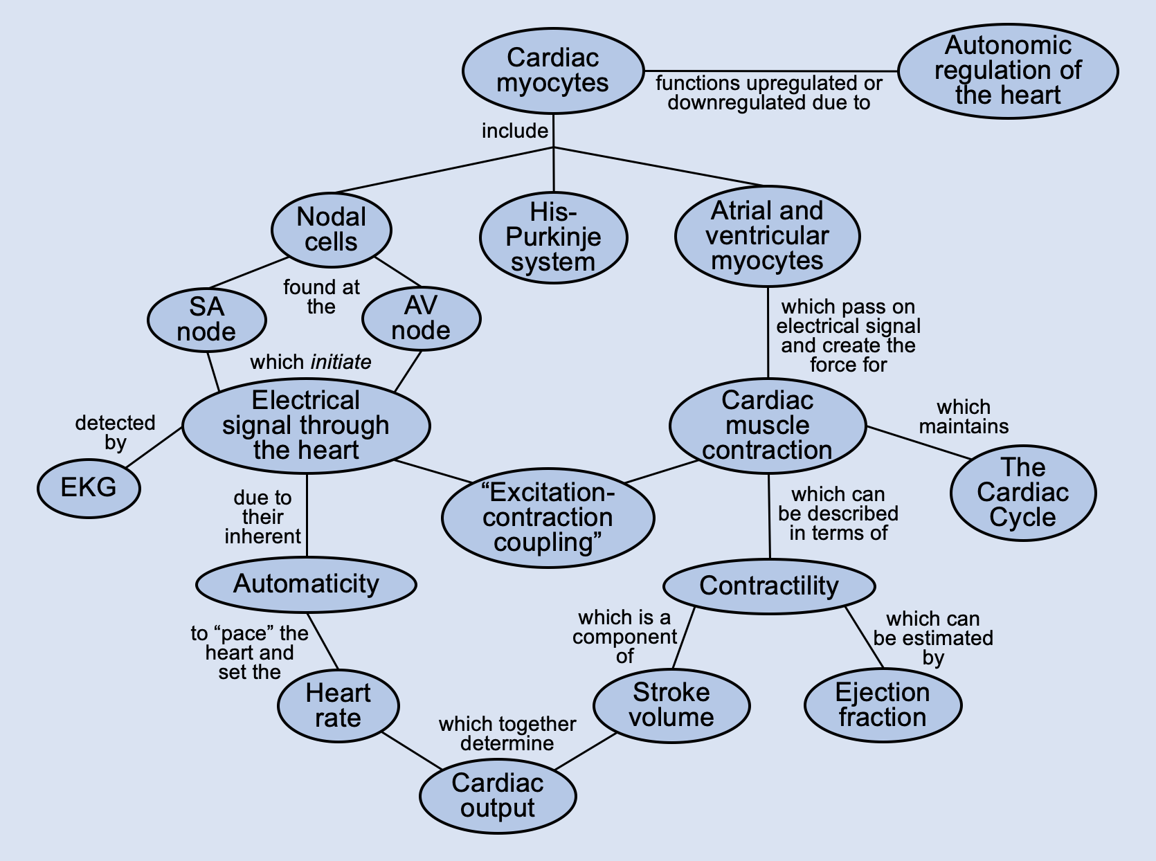
Automaticity of Cardiac Cells

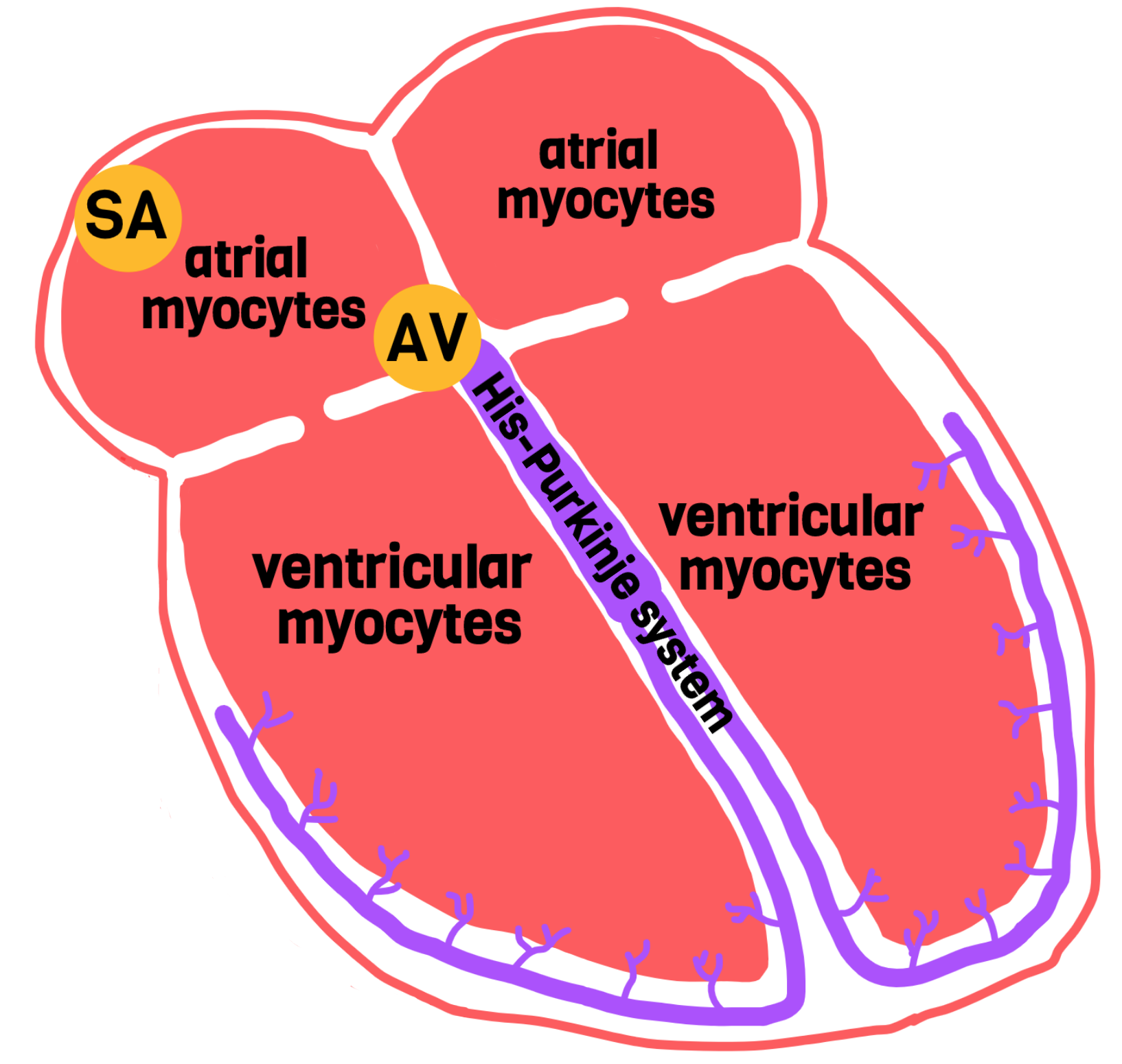
"Automaticity" refers to the ability of cardiac cells to spontaneously generate their own action potentials. In other words, they do not have to wait for a nearby cell to send it a signal in order to depolarize--they can depolarize on their own. SA nodal cells, AV nodal cells, and cells of the His-Purkinje system can all generate their own action potentials. However, they all have different firing rates.

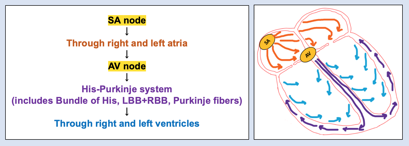
The SA node has the fastest rate of spontaneous depolarization. This means that the SA node reaches its firing threshold more quickly than the AV node or His-Purkinje cells. So, under normal circumstances, the SA node will go off before the AV node ever reaches its firing threshold, and this will start the wave of depolarization throughout the heart. Therefore, the AV node doesn't need to use its automaticity function under normal circumstances: it will always just receive the signal traveling from the SA node first. This is why the SA node is considered the main pacemaker of the heart--it sets our resting heart rate by firing about once per second. Our normal resting heart rate is the typical firing rate of the SA node (60-90 beats per minute).

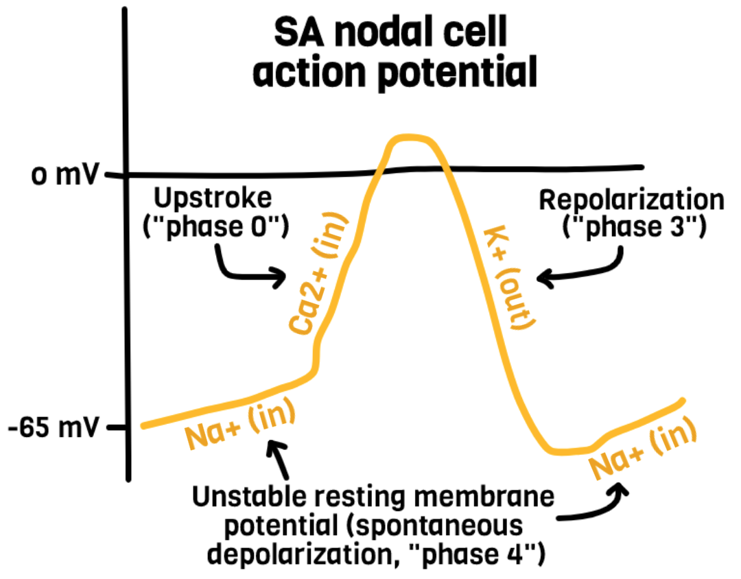
However, there are times when the SA node doesn't fire, or when the SA node's signal never reaches the AV node (eg, certain arrhythmias). In these situations, the AV node is not receiving any signal to depolarize from the SA node/atria. Thus, the AV node will just sit there, waiting to receive a signal; as the seconds tick on, the AV node's unstable resting membrane potential will finally reach its firing threshold and the AV node will depolarize on its own. This is essentially a back-up system for our heart: if something goes wrong with the SA node or the conduction in the first half of a heart beat gets messed up, the AV node has an escape route and can take over on its own. This way, our ventricles can still contract and keep our blood moving. This is why the AV node is considered the back-up pacemaker of the heart. However, whenever someone's AV node has to take over, they will likely present with bradycardia, since the automatic firing rate of the AV node is 40-60 beats per minute.