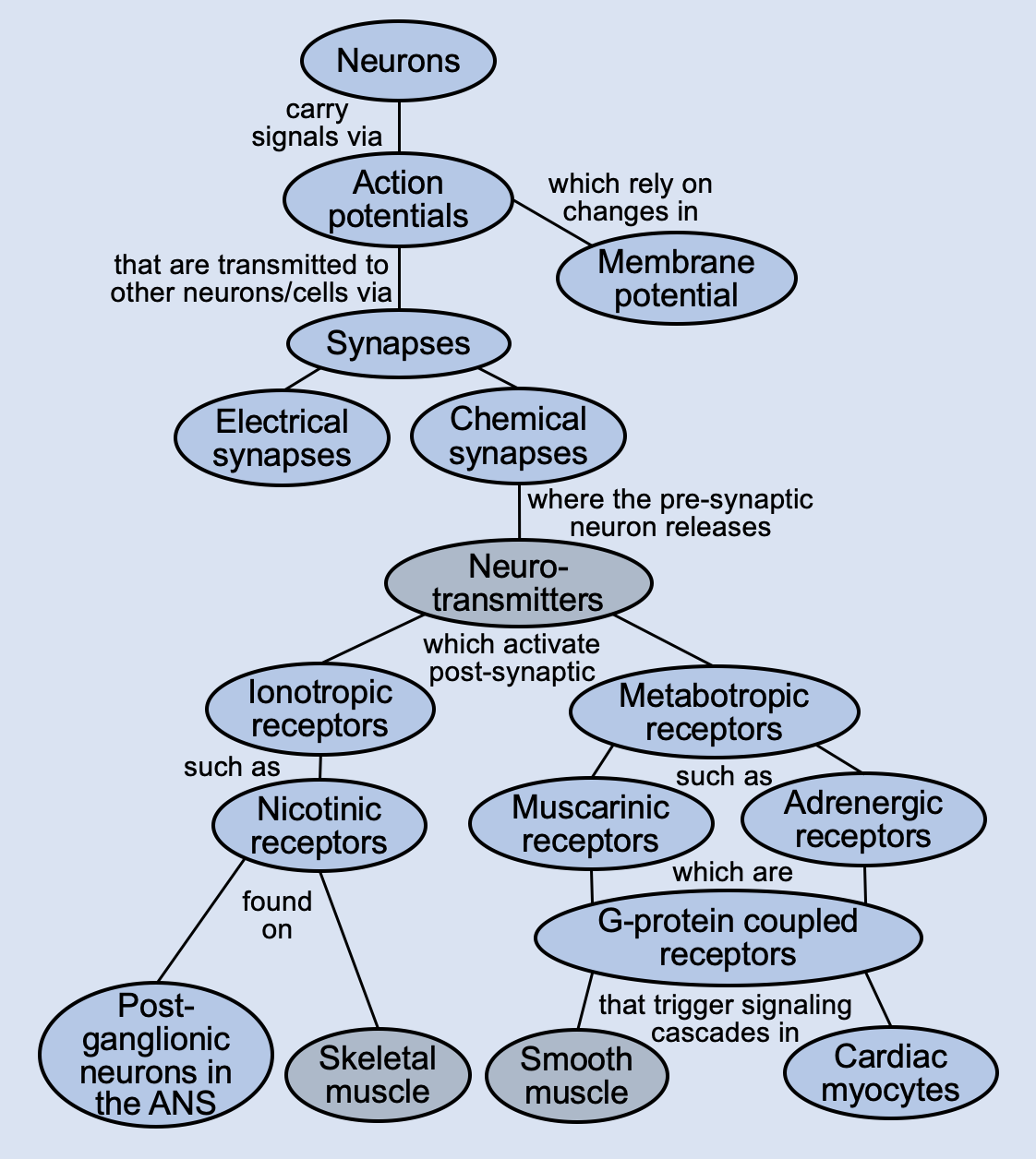
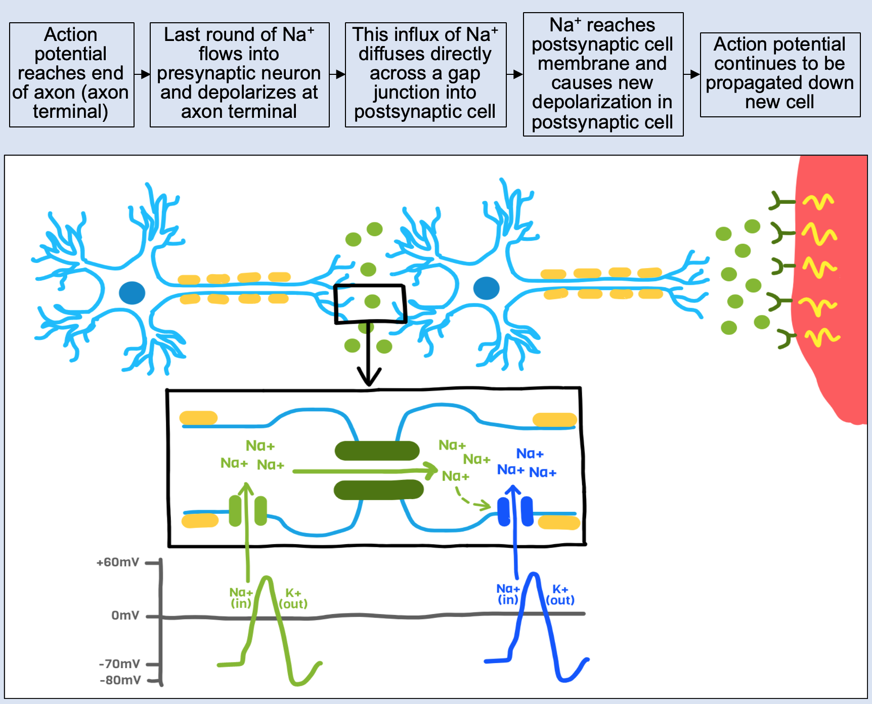
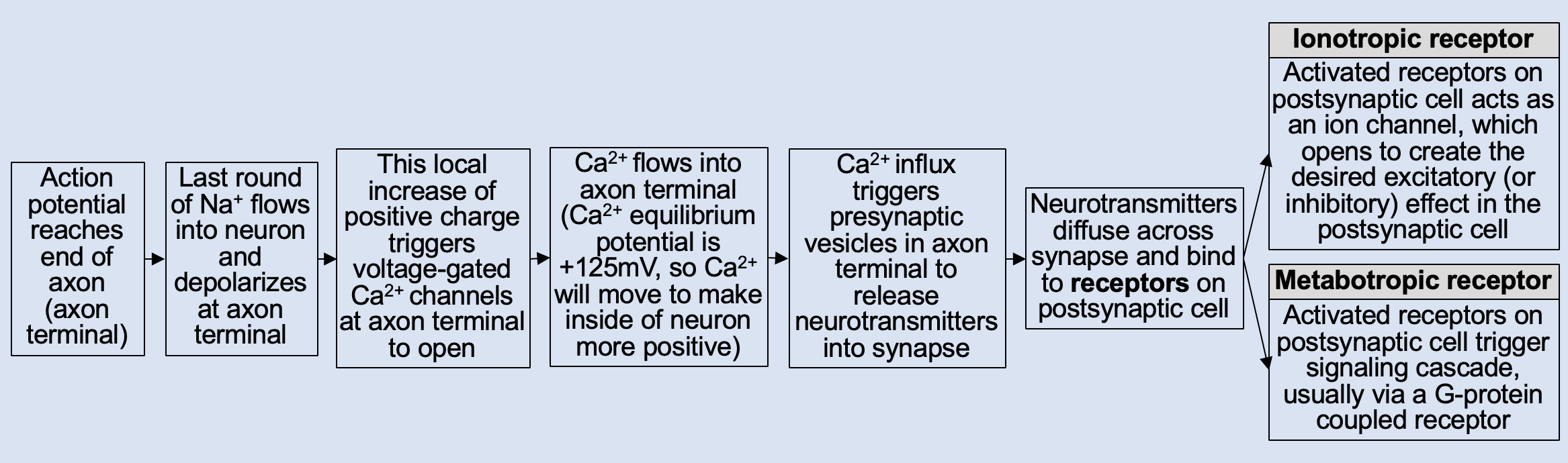
PhysiologyMaps Home

Neurons

Neurons—also called nerve cells—are the functional cell of the nervous system. The main function of neurons is to transmit signals, either a.) sensory information from the periphery to the central nervous system (via afferent, or sensory, pathways), or b.) motor information from the central nervous system to the periphery (via efferent, or motor, pathways).


A single neuron is made up of a few characteristic structures: 1.) a cell body, 2.) dendrites, 3.) an axon, and 4.) an axon terminal.


Neurons main map

Neuronal Action Potentials

Neurons transmit signals by receiving and passing on (propagating) action potentials. Neuronal action potentials are rapid movements of ions across cell membranes that can be "passed along" down the length of an axon, resulting in transmission of electrical signal. In neurons, action potentials are triggered when dendrites receive some signal from a nearby neuron. Action potentials must travel down the entire length of a neuron’s axon, in order to pass on signal to the next neuron (or to the organ it innervates).

Action potentials are created and propagated by the local movements of ions across cell membranes.

Note: While this is the characteristic action potential for a neuron, the action potentials for cardiac nodal cells, for cardiac atrial and ventricular myocytes, and for skeletal muscle cells have slightly different features.


Neurons main map

Synapses

Whenever a neuron passes on its signal to another cell, it does so at a synapse (a space or communication point between the nerve cell and another cell). The cell before the synapse (that is releasing signal from its axon terminal into the synapse) is the presynaptic cell. The cell after the synapse (that has receptors that will receive the signal from the first cell) is the postsynaptic cell. A neuron can synapse on another neuron, or a neuron can synapse on an effector organ.

There are two main types of synapses between a neuron and another neuron/cell:

  1. Electrical synapses, where ions (electrical current) are passed from one cell to the next, or
  2. Chemical synapses, where neurotransmitters cross from one cell to the next, which activate receptors on the postsynaptic cell. There are two main types of postsynaptic receptors that can be activated by neurotransmitters after crossing a chemical synapse:
    • Ionotropic receptors, which directly open ion channels in the postsynaptic cell, or
    • Metabotropic receptors, which trigger another signaling cascade (ie, a second messenger system) in the postsynaptic cell

There are two main types of effects that can be triggered when the postsynaptic receptors are activated:

  1. Excitatory responses, or
  2. Inhibitory responses.

Whether a response in excitatory or inhibitory depends on the ion channels that are affected, the neurotransmitter that is released, and/or the postsynaptic receptor that is activated.

In summary:


Neurons main map

Electrical Synapses

At electrical synapses, the signal that is passed from one cell to the next is composed of ions (electrical current). When the action potential of the presynaptic neuron reaches the axon terminal, ions diffuse directly across a gap junction into the postsynaptic cell. This flow of ions into the postsynaptic cell triggers an action potential in that cell.

Electrical synapses are typically seen:

  1. between two neurons (as depicted above), or
  2. between cardiac myocytes, via gap junctions


Neurons main map

Chemical Synapses

At chemical synapses, the signal that is passed from one cell to the next is composed of neurotransmitters, which are compounds that activate receptors in order to achieve their effects. When the action potential reaches the axon terminal of the presynaptic neuron, neurotransmitters are released from the axon terminal, diffuse into the synaptic cleft, and bind to receptors on the postsynaptic cell. This act of neurotransmitter binding to its receptor on the other side of the synapse triggers a response in the postsynaptic cell.

Neurotransmitters can trigger different physiologic responses in the postsynaptic cell by activating either:

  1. ionotropic receptors, which directly open ion channels, or
  2. metabotropic receptors, which trigger another signaling cascade, or second messenger system.


Neurons main map

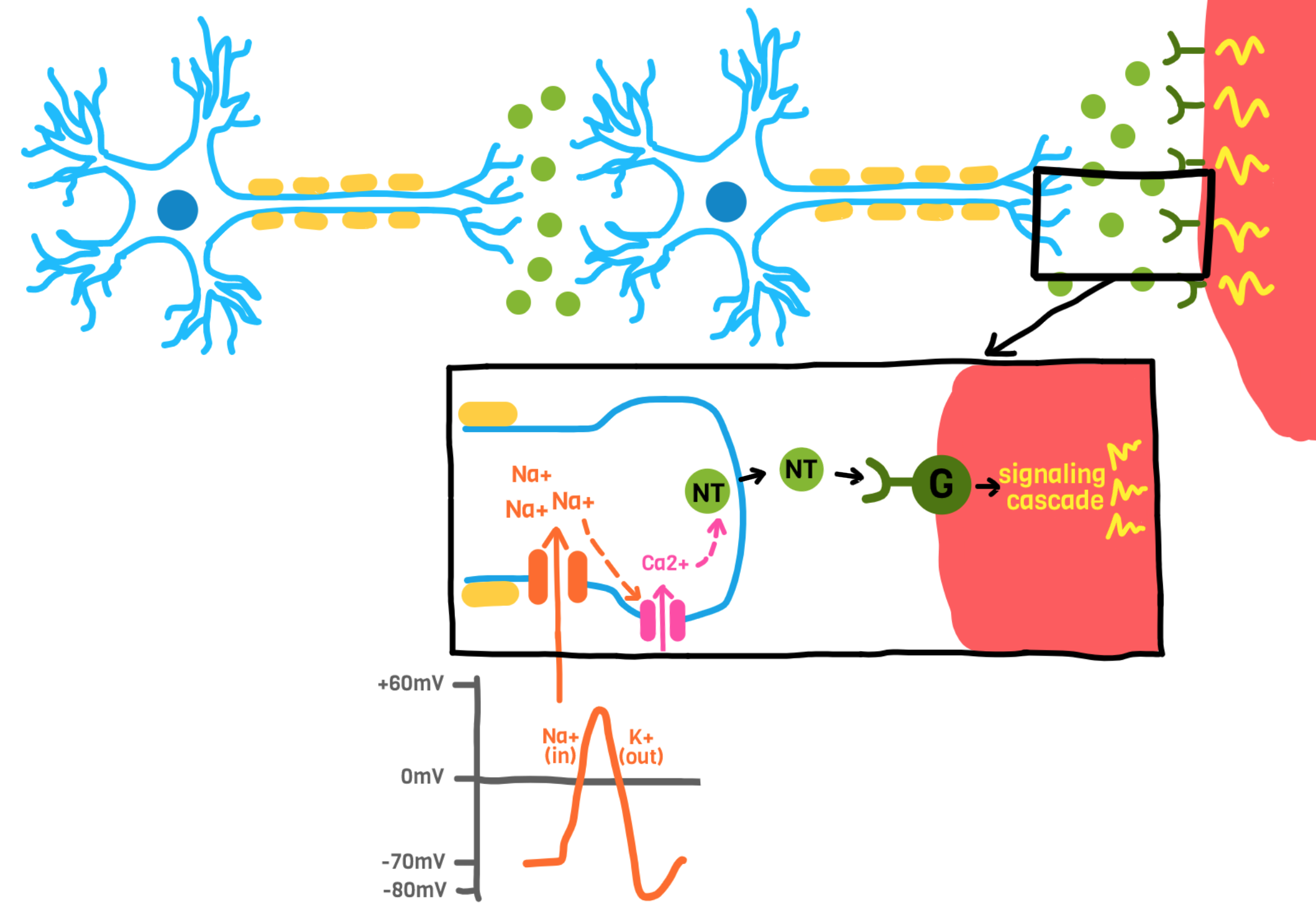
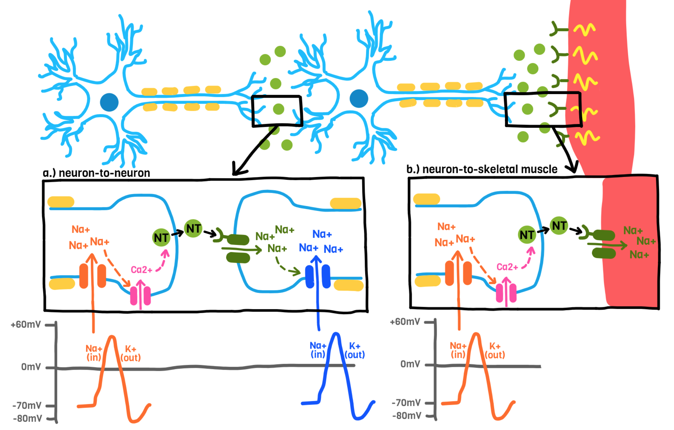
Ionotropic Receptors (at Chemical Synapses)

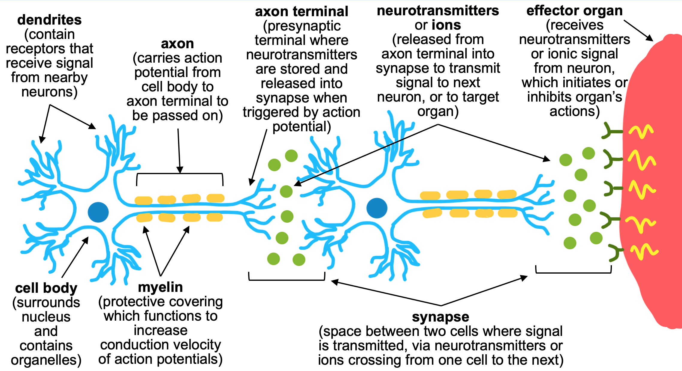
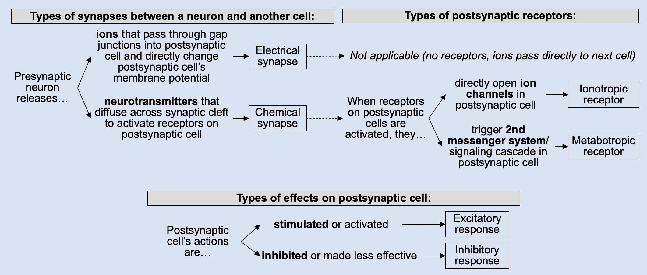
Ionotropic receptors, when activated by a neurotransmitter, directly open ion channels on the membrane of the postsynaptic cell. These are most commonly found a.) between two neurons, or b.) between a neuron and skeletal muscle (at the neuromuscular junction). Ionotropic receptors can open ion channels in the postsynaptic cell to create either an excitatory response or inhibitory response.

Excitatory Responses of Ionotropic Receptors

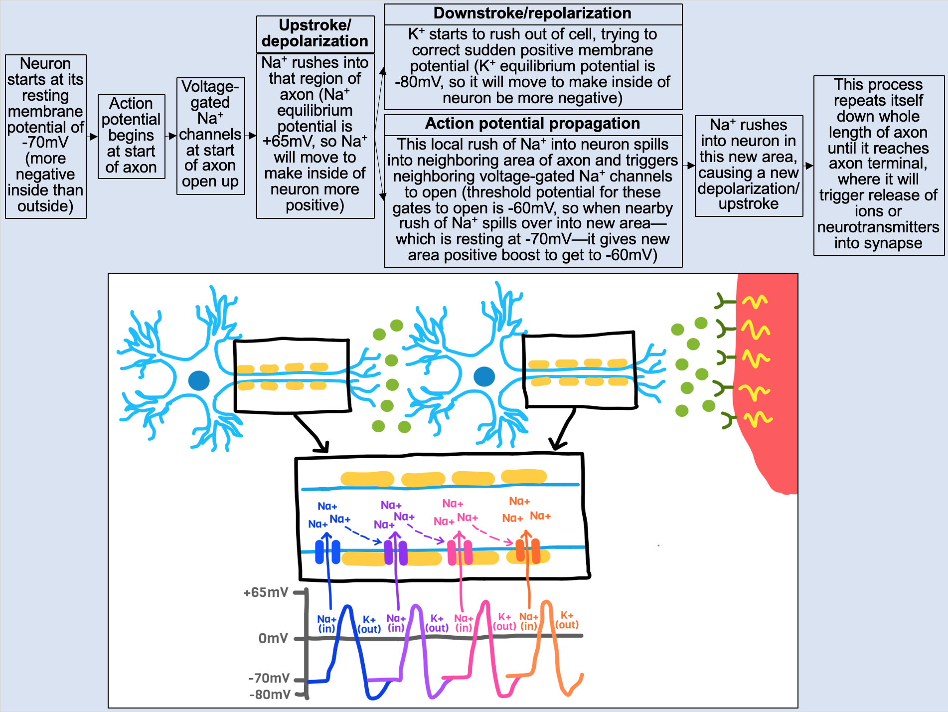
Ionotropic receptors create an excitatory response when they open Na+ channels on the postsynaptic membrane (depicted in both examples above). In both of the depicted cases, the binding of neurotransmitter at the postsynaptic cell opens Na+/K+ channels, Na+ rushes into the postsynaptic cell, and it causes a depolarization (an excitatory response, because it helped the cell get closer to its depolarization threshold). In neurons, this begins a new action potential. In skeletal muscle, this depolarization begins a muscle contraction.

Some examples of excitatory ionotropic receptors include:

Inhibitory Responses of Ionotropic Receptors

Ionotropic receptors create an inhibitory response when they open Cl- channels on the postsynaptic membrane (not depicted). When Cl- channels are opened, Cl- rushes into the postsynaptic cell (Cl- has an equilibrium potential of about -75mL, so it will want to move to make the inside of the cell more negative). This makes the postsynaptic cell's membrane potential more negative and farther away from its depolarization threshold (an inhibitory response, because it is now harder for the cell to depolarize).

Some examples of inhibitory ionotropic receptors include:

  • GABA(A) receptors in the central nervous system
  • Glycine receptors in the central nervous system

Neurons main map

Metabotropic Receptors (at Chemical Synapses)

Metabotropic receptors, when activated by a neurotransmitter, trigger another signaling cascade or second messenger system in the postsynaptic cell. This second messenger system consists of a chain of reactions that ultimately lead to the desired physiologic effect. The most common examples of metabotropic receptors are G-protein coupled receptors (GPCRs), which are some of the main receptors found in organs regulated by the autonomic nervous system.

Some examples of metabotropic receptors are:

  • Muscarinic receptors (M1, M2, M3), which are activated by acetylcholine in the autonomic nervous system
  • Adrenergic receptors (alpha-1, alpha-2, beta-1, and beta-2), which are activated by norepinephrine in the autonomic nervous system

Excitatory Responses of Metabotropic Receptors

Metabotropic receptors create an excitatory response if they trigger a stimulatory G-protein coupled receptor that activates an enzyme or increases a downstream product. Some examples of metabotropic receptors that trigger a stimulatory signaling cascade are:

  • M1 muscarinic receptors (activate the Gq pathway to increase IP3/DAG)
  • M3 muscarinic receptors (activate the Gq pathway to increase IP3/DAG)
  • alpha-1 adrenergic receptors (activate the Gq pathway to increase IP3/DAG)
  • beta-1 adrenergic receptors (activate the Gs pathway to increase cAMP)
  • beta-2 adrenergic receptors (activate the Gs pathway to increase cAMP)

Inhibitory Responses of Metabotropic Receptors

Metabotropic receptors create an inhibitory response if they trigger an inhibitory G-protein coupled receptor that inhibits some enzyme or decreases some downstream product. Some examples of metabotropic receptors that trigger an inhibitory signaling cascade are:

  • M2 muscarinic receptors (activate the Gi pathway to decrease cAMP)
  • alpha-2 adrenergic receptors (activate the Gi pathway to decrease cAMP)


Neurons main map

Effector Organs

Signal carried by neurons can trigger activity at effector organs, or organs throughout the body in which some neuronal signal has some effect. The three broad categories of effector organs, which encompass much of the functional tissue in our body, include

1.) Skeletal muscle: Neurons in the somatic nervous system synapse onto skeletal muscle at the neuromuscular junction; neurons release acetylcholine, which activate nicotinic receptors on skeletal muscle to create contractions to move our voluntary muscles,

2.) Smooth muscle: Neurons in the autonomic nervous system synapse on various types of smooth muscle throughout our body, carrying parasympathetic signals to "rest and digest" (via activation of muscarinic receptors) and sympathetic signals to initiate "fight or flight" (via activation of adrenergic receptors), and

3.) Cardiac muscle: The heart is under tight autonomic regulation, so it also receives signals from autonomic nervous system neurons to initiate "rest" (parasympathetic effects, downregulating heart activity via muscarinic receptors) and "fight or flight" (sympathetic effects, upregulating heart activity via adrenergic receptors).