Autonomic Nervous System (ANS)
Click here for an overview of neurons, neuronal action potentials, and synapses
Click here to jump down to the ANS summary chart
Autonomic Nervous System (ANS)
Click here for an overview of neurons, neuronal action potentials, and synapses Click here to jump down to the ANS summary chart |
|
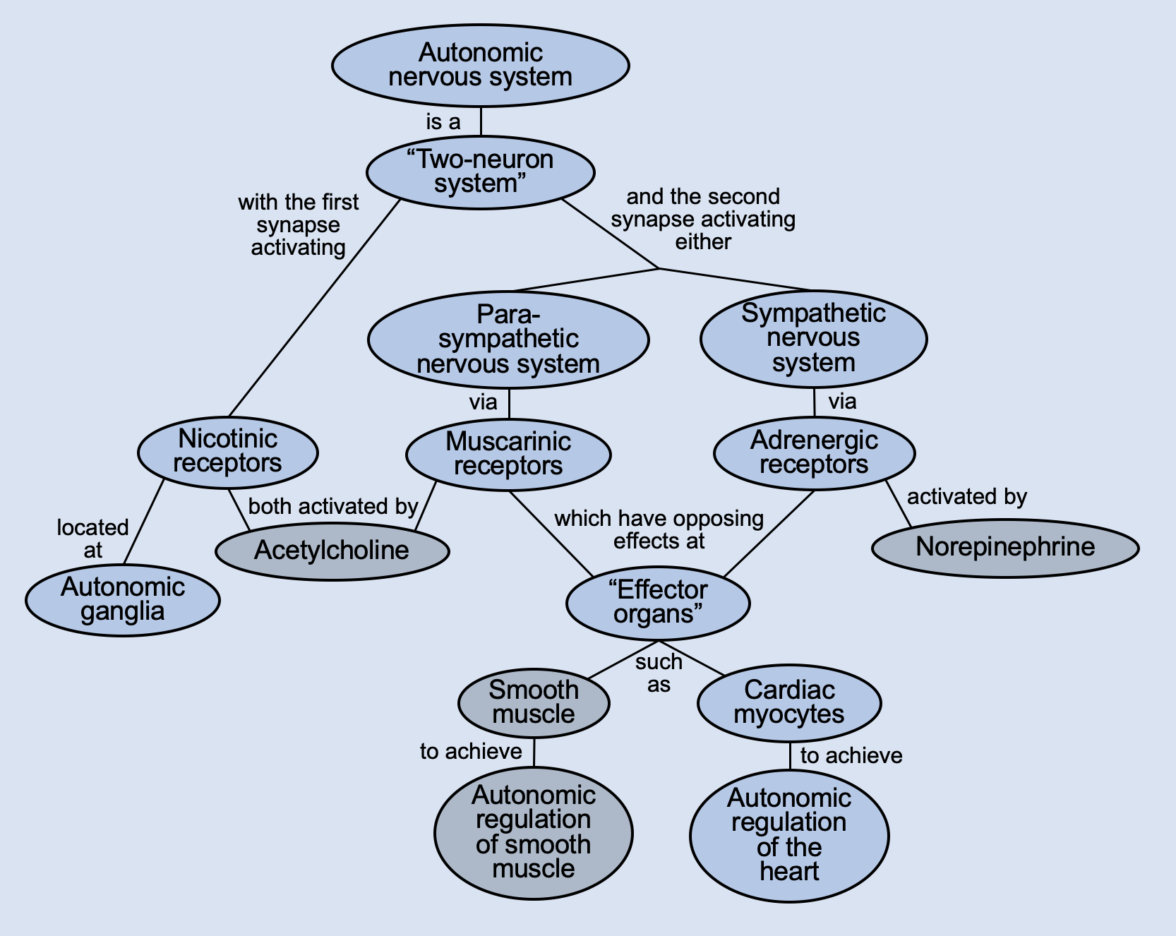
PhysiologyMaps Home > Autonomic Nervous System The ANS is a Two-Neuron SystemThe autonomic nervous system is a two-neuron system (in contrast to the somatic nervous system, which is a one-neuron system). This means that anytime our brain tells the ANS to do something, that signal always travels through two neurons to get to its site of action. Signal travels through this 2-neuron system via neuronal action potentials. Neuron 1 originates in the central nervous system (CNS) and always releases the neurotransmitter acetylcholine (ACh) into its synapse (which activates nicotinic receptors on Neuron 2). Neuron 1 synapses onto Neuron 2 at an autonomic ganglion, which are basically just dedicated areas in our body where neurons synapse on each other. Thus, Neuron 1 is called the preganglionic neuron (before the ganglia) and Neuron 2 is called the postganglionic neuron (after the ganglia). Neuron 2 then carries signal to the organ we are trying to regulate ("effector organ"), where it triggers a G protein-coupled receptor that will activate a signaling cascade to achieve specific effects in that organ. Neuron 2's neurotransmitters, the receptors it activates, and the G protein subtype it triggers all depend on whether we are dealing with the parasympathetic nervous system or the sympathetic nervous system. 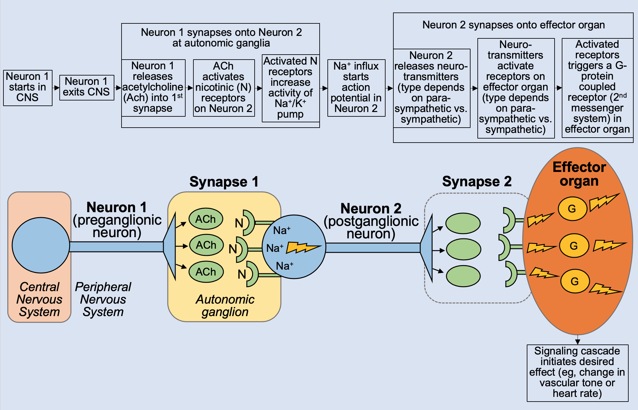 Nicotinic Receptors in the ANSNicotinic receptors in the ANS are found on the postganglionic neuron (the start of the 2nd neuron in the 2-neuron chain). Nicotinic receptors are activated by acetylcholine traveling from Neuron 1 to Neuron 2. Once activated, they directly open Na+ channels in Neuron 2, which starts Neuron 2's action potential, allowing the neuronal signal to propagate toward the effector organ. Nicotinic receptors are an example of an ionotropic receptor, since they directly open an ion channel to achieve their effects. Note: Nicotinic receptors can also be found on skeletal muscle (where a neuron releases acetylcholine into the neuromuscular junction to initiate voluntary movement). However, this is considered part of the somatic nervous system, not the autonomic nervous system. |
|
PhysiologyMaps Home > Autonomic Nervous System ANS Effector OrgansThere are two main groups of organs that are regulated by the autonomic nervous system: 1.) smooth muscle and 2.) the heart. They are called "effector organs" because they are the organs where we want to create some autonomic effect that will help us either "rest and digest" (parasympathetic effects) or "fight or flight" (sympathetic effects). Smooth muscle is found in many different organs of our body, including blood vessels (to constrict or dilate), the GI tract (to increase or decrease motility), the eye (to dilate or constrict the pupil), the bladder wall, and GI and bladder sphincters. The parasympathetic and sympathetic divisions of the ANS affect these smooth muscles in different ways to help us achieve our normal bodily functions; these effects are detailed below. The heart is also under tight autonomic regulation. The ANS controls our heart rate (chronotropic effects), the contractility of our cardiac myocytes (inotropic effects), and the conduction velocity of electrical signal through the heart (dromotropic effects). The parasympathetic and sympathetic divisions of the ANS have oppsite effects on these outcomes (with parasympathetic usually slowing down our heart functions, and sympathetic jacking up our heart functions). |
|
PhysiologyMaps Home > Autonomic Nervous System Autonomic Ganglia***. |
|
PhysiologyMaps Home > Autonomic Nervous System Parasympathetic Nervous SystemThe parasympathetic division of the ANS initiates "rest and digest" activities, such as increased GI motility, urination, and decreased heart rate. To achieve these effects, the postganglionic neurons of the parasympathetic nervous system release the neurotransmitter acetylcholine, which activates muscarinic receptors on effector organs (in contrast to preganglionic neurons, which also release acetylcholine but activate nicotinic receptors.) 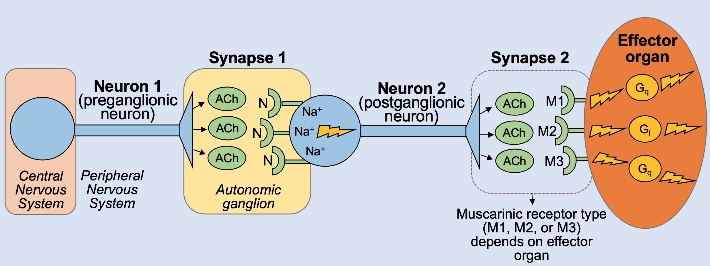 Muscarinic Receptors in the ANSMuscarinic receptors are found on many organs organs throughout the body and are activated by acetylcholine released by parasympathetic nervous system neurons. The main function of activated muscarinic receptors is to trigger the "rest and digest" effects of the parasympathetic nervous system (with one exception). To do this, activated muscarinic receptors trigger different G protein-coupled receptor cascades, which then go on to activate (or inhibit) organ functions. The three main subtypes of muscarinic receptors are M1, M2, and M3. Their locations, associated G-protein systems, and effects are summarized in the table below. 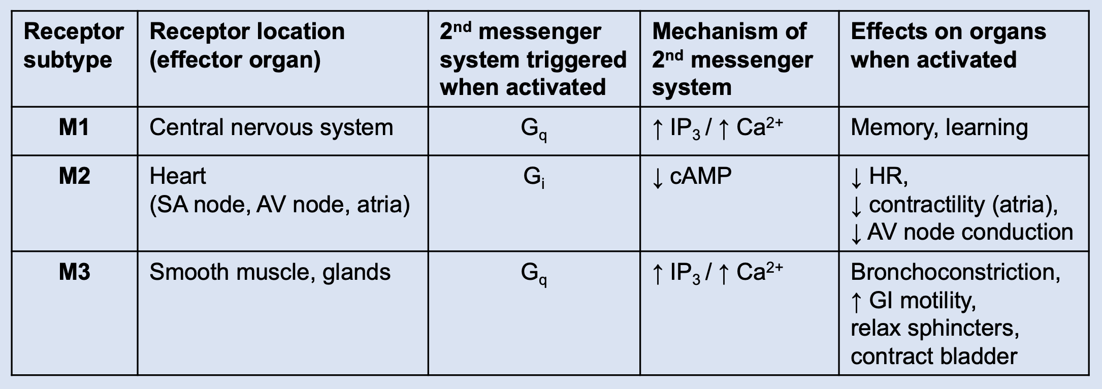 One exception to the "parasympathetic-muscarinic" match-up is the muscarinic receptors on sweat glands, which are activated by acetylcholine released from sympathetic nervous system neurons (not by parasympathetic neurons). |
|
PhysiologyMaps Home > Autonomic Nervous System Sympathetic Nervous SystemThe sympathetic division of the ANS initiates "fight or flight" activities, such as increased heart rate, increased heart contractility, and bronchodilation. To achieve these effects, the postganglionic neurons of the sympathetic nervous system mainly release the neurotransmitter norepinephrine (with one exception), which activates adrenergic receptors on effector organs. The one exception to the "sympathetic-adrenergic" match-up is the sympathetic activation of sweat glands; in this case, the sympathetic neuron releases acetylcholine (instead of norepinephrine), which activates muscarinic receptors (instead of adrenergic receptors). 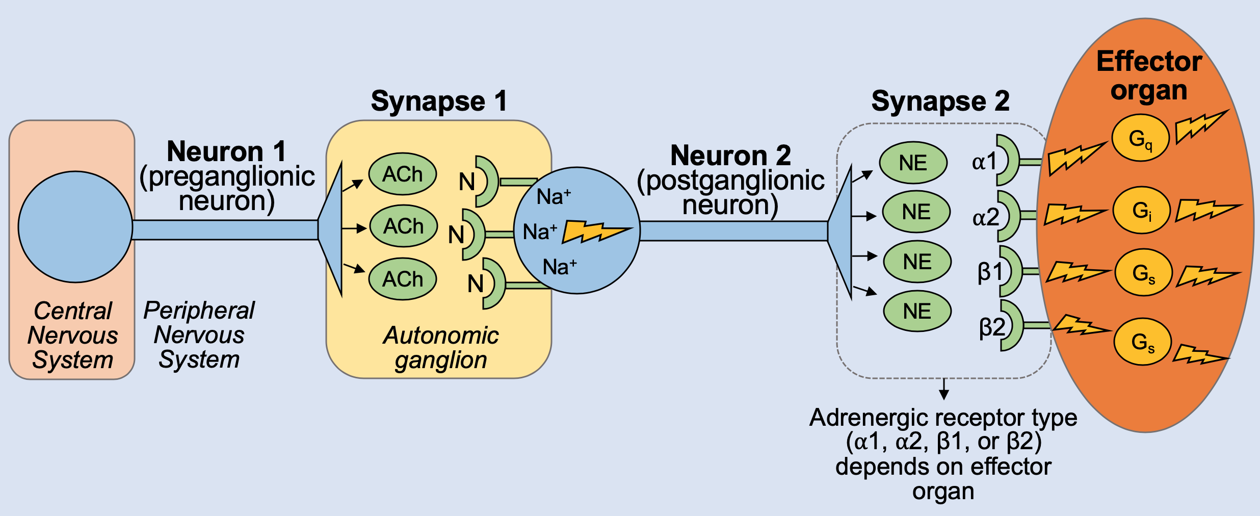 Adrenergic Receptors in the ANSAdrenergic receptors are found on many organs organs throughout the body and are activated by norepinephrine released by sympathetic nervous system neurons. The main function of activated adrenergic receptors is to trigger the "fight or flight" effects the sympathetic nervous system. To do this, adrenergic receptors trigger different G protein-coupled receptor cascades, which then go on to activate (or inhibit) organ functions. The four main subtypes of adrenergic receptors are alpha-1, alpha-2, beta-1, and beta-2. Their locations, associated G-protein systems, and effects are summarized in the table below. 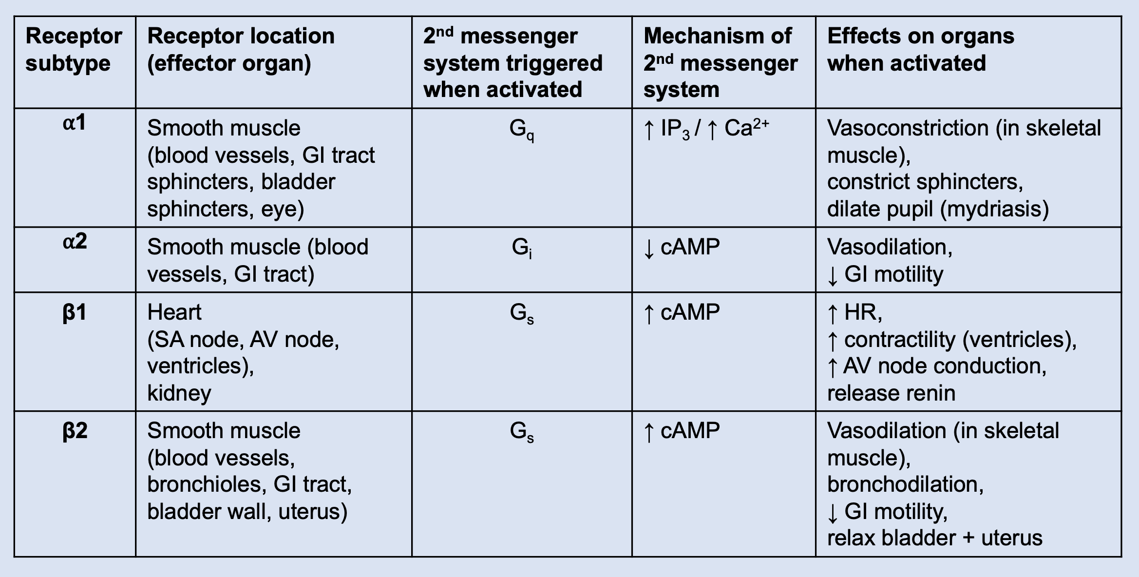 |
|
PhysiologyMaps Home > Autonomic Nervous System Autonomic Nervous System: Summary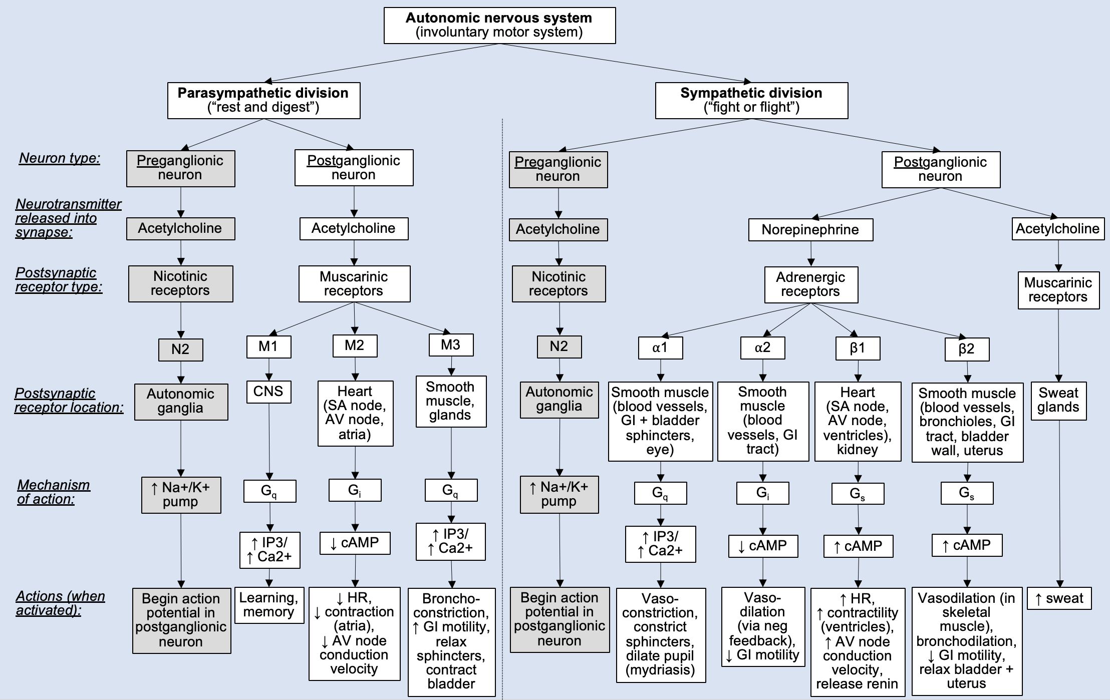 |