|
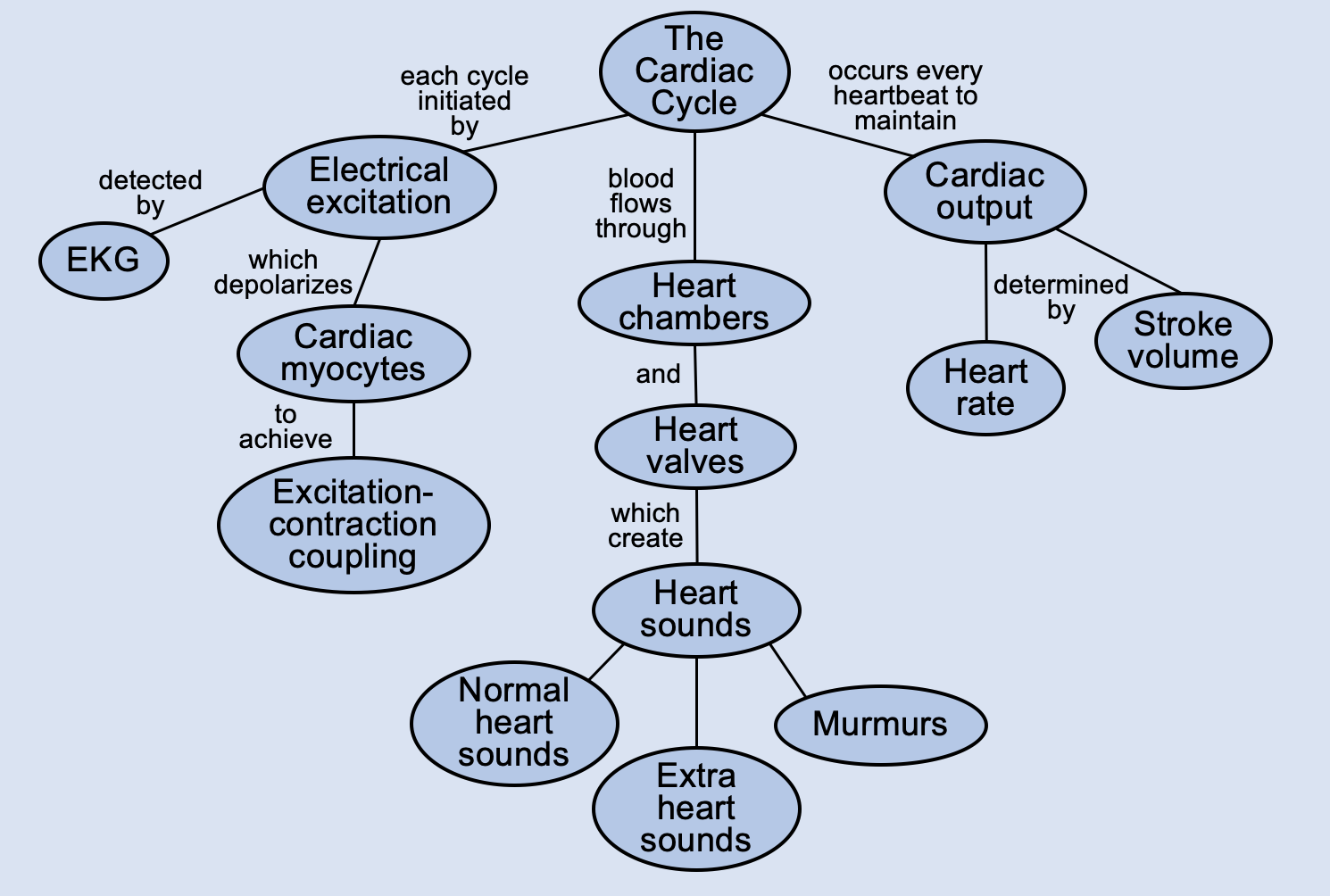
PhysiologyMaps Home > Cardiovascular main > The Cardiac Cycle
Extra Heart Sounds (S3, S4, and splitting)
Sometimes, we can hear extra heart sounds on auscultation. Sometimes these can occur in normal hearts, but they sometimes indicate disesae.
S3
S3 occurs in early diastole (right after S2) when there is really rapid or forceful filling of the LV (the S3 sound is essentially blood slamming against the LV wall during early filling, right after the mitral valve opens). This usually happens in the context of a high LA pressure, which leads to a swift rush of blood from the LA to the LV during diastole. An S3 can be present in the following situations:
- Mitral regurgitation: xxx.
- Acute heart failure: xxx.
- Can be normal in young patients and pregnant women: xx.
S4
S4 occurs in late diastole (right before the next S1) when the LV pressures are so high that the LA needs to give a little extra contraction at the end of diastole in order to empty all its blood into the high-pressure LV (this is why it is called an "atrial kick"). This usually happens in the context of a stiff LV, which creates a high-pressure LV even during diastole, when it should be more relaxed. An S4 can be present in the following situations:
- LV hypertrophy: xxx.
- Hypertrophic cardiomyopathy: xxx.
- Diastolic heart failure: xxx.
Note: Left-sided S3 and S4 are associated with left heart abnormalities. Right-sided S3 and S4 are associated with right heart abnormalities.
Splitting of S2
The second heart sound (S2) can sometimes seem to be "split" into two separate sounds. S2 is normally created by the simultaneous closure of the pulmonic and aortic valves. So, S2 splitting occurs if either the pulmonic or aortic valve closure is delayed, so that the pulmonic valve and aortic valve do not close at the same time.
- Normal delay of pulmonic valve closure (normal S2 splitting): xxx.
- Abnormal delay of pulmonic valve closure: xxx.
- Abnormal delay of aortic valve closure: xxx.
Other sounds that can be heard during auscultation include heart murmurs, which usually signify diseases or abnormalities of heart valves or blood flow.
|