|
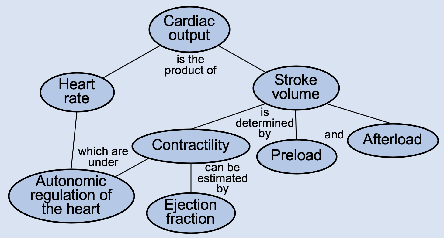
PhysiologyMaps Home > Cardiovascular main > Cardiac output
Ejection Fraction
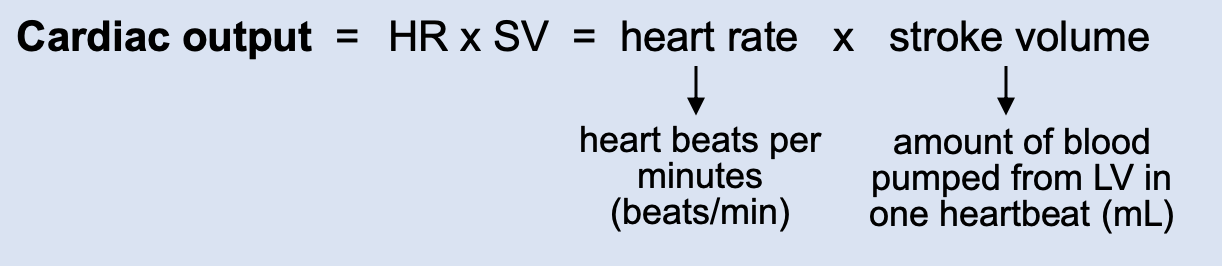
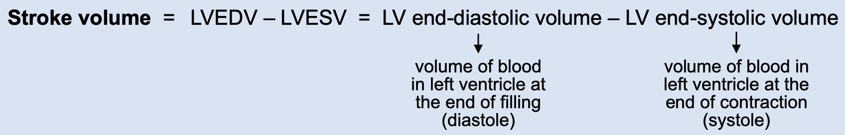
Ejection fraction (EF) is the percentage of blood in a filled left ventricle (LV) that is pumped out to the body in one heartbeat. The LV does not typically pump out 100% of the blood that it contains at the end of diastole (ventricular filling). A normal EF is around 55-70%, meaning that the left ventricle only pumps out about 55-70% of the blood it contains with each heartbeat. Ejection fraction can be expressed as:
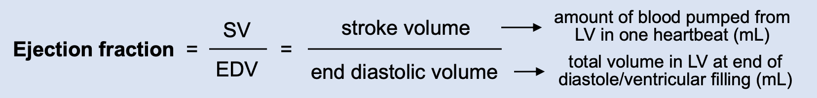
EF can be used as an estimation of the contractility of the cardiac muscle, or the force the cardiac myocytes generate during each contraction. If contractility of the LV increases, the EF will increase, because the LV will be able to generate more force to pump out more blood during systole, given a constant starting volume.
Ejection Fraction and Heart Failure
In important clinical correlate of EF is when it is used to describe heart failure. In general, heart failure is characterized by a decrease in cardiac output. However, there are two kinds of heart failure, which depend on whether EF is reduced or not reduced (preserved):
- Heart failure with reduced EF (HFrEF): This refers to "systolic heart failure (HF)". When a person has HF with systolic dysfunction, it means that they have a problem pumping blood out of the LV. Let's say that in a normal heartbeat, the LV gets filled with 100mL of blood by the end of diastole and pumps out 60mL of blood during systole (so the EDV is 100mL, and the SV is 60mL). This gives us a normal EF of 60%. In systolic HF, the problem is with pumping--so we will fill normally (EDV of 100mL), but we won't pump normally (we have SV of, let's say, 40mL). This will certainly decrease our cardiac output (because we went from pumping out 60mL of blood every heartbeat to just 40mL), so we will be diagnosed with HF. Our new EF will be 40% (40mL/100mL), which is below the normal limit of 55-70%, so we will have "HF with reduced EF".
- Heart failure with preserved EF (HFpEF): This refers to "diastolic heart failure (HF)". When a person has heart failure with diastolic dysfunction, it means that their LV can pump fine, but they have a problem with filling the ventricle. Let's again say that in a normal heartbeat, the LV gets filled with 100mL of blood by the end of diastole and pumps out 60mL of blood during systole (so the EDV is 100mL, and the SV is 60mL, giving us a normal EF of 60%). In diastolic HF, we won't fill normally during diastole (we have an EDV of, let's say 70mL, instead of our normal 100mL). We don't have any problem with pumping during systole, so for every heartbeat, we still pump out about 60% of our LV's blood volume. But 60% of 70mL is only around 40mL (so our SV for each heartbeat is only 40mL). This will certainly decrease our cardiac output (because we went from pumping out 60mL of blood for every heartbeat to just 40mL), so we will be diagnosed with HF. However, 40mL/70mL gives us an EF of 60%, which is normal, so we will have "HF with preserved EF".
|